
What is a pacemaker?
A pacemaker is composed of three parts: a pulse generator, one or more leads, and an electrode on each lead. A pacemaker signals the heart to beat when the heartbeat is too slow or irregular.
A pulse generator is a small metal case that contains electronic circuitry with a small computer and a battery that regulate the impulses sent to the heart.
The lead (or leads) is an insulated wire that is connected to the pulse generator on one end, with the other end placed inside one of the heart’s chambers. The lead is almost always placed so that it runs through a large vein in the chest leading directly to the heart.
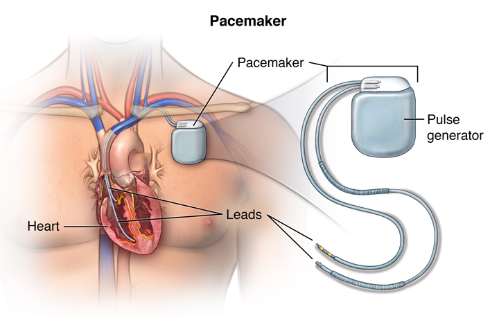
The electrode on the end of a lead touches the heart wall. The lead delivers the electrical impulses to the heart. It also senses the heart’s electrical activity and relays this information back to the pulse generator. Pacemaker leads may be positioned in the atrium (upper chamber) or ventricle (lower chamber) or both, depending on the medical condition.
If the heart’s rate is slower than the programmed limit, an electrical impulse is sent through the lead to the electrode and causes the heart to beat at a faster rate.
When the heart beats at a rate faster than the programmed limit, the pacemaker generally monitors the heart rate and will not pace. Modern pacemakers are programmed to work on demand only, so they do not compete with natural heartbeats. Generally, no electrical impulses will be sent to the heart unless the heart’s natural rate falls below the pacemaker’s lower limit.
A newer type of pacemaker, called a biventricular pacemaker, is currently used in the treatment of specific types of heart failure. Sometimes in heart failure, the two ventricles do not pump in a normal manner. Ventricular dyssynchrony is a common term used to describe this abnormal pumping pattern. When this happens, less blood is pumped by the heart. A biventricular pacemaker paces both ventricles at the same time, increasing the amount of blood pumped by the heart. This type of treatment is called cardiac resynchronization therapy or CRT.
After a pacemaker insertion, regularly scheduled appointments will be made to ensure the pacemaker is functioning properly. The doctor uses a special computer, called a programmer, to review the pacemaker’s activity and adjust the settings when needed.
Other related procedures that may be used to assess the heart include resting and exercise electrocardiogram (ECG), Holter monitor, signal-averaged ECG, cardiac catheterization, chest X-ray, computed tomography (CT scan) of the chest, echocardiography, electrophysiology studies, magnetic resonance imaging (MRI) of the heart, myocardial perfusion scan (stress), myocardial perfusion scan (resting), radionuclide angiography, and cardiac CT scan. Please see these procedures for additional information. Note that although an MRI is a very safe procedure, the magnetic fields used by the MRI scanner may interfere with the pacemaker’s function. Any patient with a pacemaker should always speak with his or her cardiologist before undergoing an MRI.
Procedure overview
What is a pacemaker insertion?
A pacemaker insertion is the implantation of a small electronic device that is usually placed in the chest (just below the collarbone) to help regulate slow electrical problems with the heart. A pacemaker may be recommended to ensure that the heartbeat does not slow to a dangerously low rate.
The heart’s electrical system
The heart is basically a pump made up of muscle tissue that is stimulated by electrical currents, which normally follow a specific circuit within the heart.
This normal electrical circuit begins in the sinus or sinoatrial (SA) node, which is a small mass of specialized tissue located in the right atrium (upper chamber) of the heart. The SA node generates an electrical stimulus at 60 to 100 times per minute (for adults) under normal conditions; this electrical impulse from the SA node starts the heartbeat.
The electrical impulse travels from the SA node via the atria to the atrioventricular (AV) node in the bottom of the right atrium. From there the impulse continues down an electrical conduction pathway called the Bundle of His and then on through the “His-Purkinje” system into the ventricles (lower chambers) of the heart. When the electrical stimulus occurs it causes the muscle to contract and pump blood to the rest of the body. This process of electrical stimulation followed by muscle contraction is what makes the heart beat.
A pacemaker may be needed when problems occur with the electrical conduction system of the heart. When the timing of the electrical stimulation of the heart to the heart muscle and the subsequent response of the heart’s pumping chambers is altered, a pacemaker may help.
Reasons for the procedure
A pacemaker may be inserted in order to stimulate a faster heart rate when the heart is beating too slowly, and causing problems that cannot otherwise be corrected.
Problems with the heart rhythm may cause difficulties because the heart is unable to pump an adequate amount of blood to the body. If the heart rate is too slow, the blood is pumped too slowly. If the heart rate is too fast or too irregular, the heart chambers are unable to fill up with enough blood to pump out with each beat. When the body does not receive enough blood, symptoms such as fatigue, dizziness, fainting, and/or chest pain may occur.
Some examples of heart rate and rhythm problems for which a pacemaker might be inserted include:
- Bradycardia. This occurs when the sinus node causes the heart to beat too slowly.
- Tachy-brady syndrome. This is characterized by alternating fast and slow heartbeats.
- Heart block. This occurs when the electrical signal is delayed or blocked after leaving the SA node; there are several types of heart blocks.
There may be other reasons for your doctor to recommend a pacemaker insertion.
Risks of the procedure
Possible risks of pacemaker include, but are not limited to, the following:
- Bleeding from the incision or catheter insertion site
- Damage to the vessel at the catheter insertion site
- Infection of the incision or catheter site
- Pneumothorax. If the nearby lung is inadvertently punctured during the procedure, leaking air becomes trapped in the pleural space (outside the lung but within the chest wall); this can cause breathing difficulties and in extreme cases may cause the lung to collapse.
If you are pregnant or suspect that you may be pregnant, you should notify your health care provider. If you are breastfeeding, you should notify your health care provider.
Patients who are allergic to or sensitive to medications or latex should notify their doctor.
For some patients, having to lie still on the procedure table for the length of the procedure may cause some discomfort or pain.
There may be other risks depending on your specific medical condition. Be sure to discuss any concerns with your doctor prior to the procedure.
Before the procedure
Your doctor will explain the procedure to you and offer you the opportunity to ask any questions that you might have about the procedure:
- You will be asked to sign a consent form that gives your permission to do the test. Read the form carefully and ask questions if something is not clear.
- Notify your doctor if you are sensitive to or are allergic to any medications, iodine, latex, tape, or anesthetic agents (local and general).
- You will need to fast for a certain period of time prior to the procedure. Your doctor will notify you how long to fast, usually overnight.
- If you are pregnant or suspect that you are pregnant, you should notify your doctor.
- Notify your doctor of all medications (prescription and over-the-counter) and herbal or other supplements that you are taking.
- Notify your doctor if you have heart valve disease, as you may need to receive an antibiotic prior to the procedure.
- Notify your doctor if you have a history of bleeding disorders or if you are taking any anticoagulant (blood-thinning) medications, aspirin, or other medications that affect blood clotting. It may be necessary for you to stop some of these medications prior to the procedure.
- Your doctor may request a blood test prior to the procedure to determine how long it takes your blood to clot. Other blood tests may be done as well.
- You may receive a sedative prior to the procedure to help you relax. If a sedative is given and there is a possibility that you may be discharged, you will need someone to drive you home. You will likely spend at least one night in the hospital after the procedure for observation and to ensure the pacemaker functions properly.
- Based on your medical condition, your doctor may request other specific preparation.
During the procedure
A pacemaker may be performed on an outpatient basis or as part of your stay in a hospital. Procedures may vary depending on your condition and your doctor’s practices.
Generally, a pacemaker insertion follows this process:
- You will be asked to remove any jewelry or other objects that may interfere with the procedure.
- You will be asked to remove your clothing and will be given a gown to wear.
- You will be asked to empty your bladder prior to the procedure.
- If there is excessive hair at the incision site, it may be clipped off.
- An intravenous (IV) line will be started in your hand or arm prior to the procedure for injection of medication and to administer IV fluids, if needed.
- You will be placed on your back on the procedure table.
- You will be connected to an electrocardiogram (ECG or EKG) monitor that records the electrical activity of the heart and monitors the heart during the procedure using small, adhesive electrodes. Your vital signs (heart rate, blood pressure, breathing rate, and oxygenation level) will be monitored during the procedure.
- Large electrode pads will be placed on the front and back of the chest.
- You will receive a sedative medication in your IV before the procedure to help you relax. However, you will likely remain awake during the procedure.
- The pacemaker insertion site will be cleansed with antiseptic soap.
- Sterile towels and a sheet will be placed around this area.
- A local anesthetic will be injected into the skin at the insertion site.
- Once the anesthetic has taken effect, the physician will make a small incision at the insertion site.
- A sheath, or introducer, is inserted into a blood vessel, usually under the collarbone. The sheath is a plastic tube through which the pacer lead wire will be inserted into the blood vessel and advanced into the heart.
- It will be very important for you to remain still during the procedure so that the catheter does not move out of place and to prevent damage to the insertion site.
- The lead wire will be inserted through the introducer into the blood vessel. The doctor will advance the lead wire through the blood vessel into the heart.
- Once the lead wire is inside the heart, it will be tested to verify proper location and that it works. There may be one, two, or three lead wires inserted, depending on the type of device your doctor has chosen for your condition. Fluoroscopy, (a special type of X-ray that will be displayed on a TV monitor), may be used to assist in testing the location of the leads.
- The pacemaker generator will be slipped under the skin through the incision (just below the collarbone) after the lead wire is attached to the generator. Generally, the generator will be placed on the nondominant side. (If you are right-handed, the device will be placed in your upper left chest. If you are left-handed, the device will be placed in your upper right chest).
- The ECG will be observed to ensure that the pacer is working correctly.
- The skin incision will be closed with sutures, adhesive strips, or a special glue.
- A sterile bandage or dressing will be applied.
After the procedure
In the hospital
After the procedure, you may be taken to the recovery room for observation or returned to your hospital room. A nurse will monitor your vital signs.
You should immediately inform your nurse if you feel any chest pain or tightness, or any other pain at the incision site.
After the period of bed rest has been completed, you may get out of bed with assistance. The nurse will assist you the first time you get up, and will check your blood pressure while you are lying in bed, sitting, and standing. You should move slowly when getting up from the bed to avoid any dizziness from the period of bedrest.
You will be able to eat or drink once you are completely awake.
The insertion site may be sore or painful. Pain medication may be administered if needed.
Your doctor will visit with you in your room while you are recovering. The doctor will give you specific instructions and answer any questions you may have.
Once your blood pressure, pulse, and breathing are stable and you are alert, you will be taken to your hospital room or discharged home.
If the procedure is performed on an outpatient basis, you may be allowed to leave after you have completed the recovery process. However, it is common to spend at least one night in the hospital after pacemaker implantation for observation.
To Get An Appointment Today (+91) 9607799333